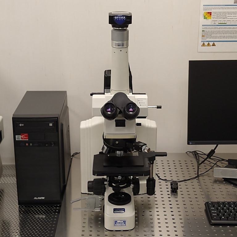
Optical Microscope – Nikon Eclipse ME600
Details
Description:
Nikon Eclipse ME600 is optical microscope equipped with five objectives (5X, 10X, 20X, 50X and 100X), featuring both reflected (epi) and transmitted (dia) illumination systems. The microscope can be used in differential interference contrast (DIC) imaging, which is an imaging method based on the contrast difference of the samples, and in dark field (DF) mode (except with 10X objective). With DIC and DF, it is possible to see details from optically transparent samples that are invisible in the ordinary microscope images. It is also equipped with a high performance 6.3 Megapixel color CMOS camera, suitable for high quality image/video capture and line/surface/angle measurements.
Working principle
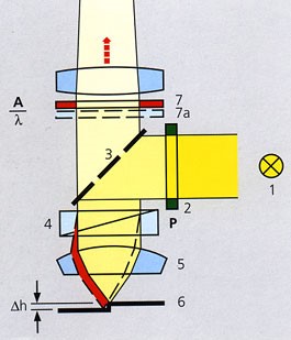
Differential Interference Contrast (DIC)
This method, an extension of polarization contrast, is suitable for the visualization of even minute differences in height on surfaces. A birefringent prism (4) is used which splits the polarized light beam into two partial beams on its way to the sample. These two partial beams strike the sample (6) with lateral displacement from each other. If the surface is completely flat, nothing will happen. However, if there is a small step between the partial rays, one of the two beams has to travel along a path which is 2Δh longer. Once the partial beams have returned via DIC prism (4) and analyzer (7), they display the same direction of vibration again and can interfere with each other in the intermediate image. The path difference experienced on the surface then changes into grey values which can be seen by the eye: steps become visible in the form of relief. As an auxiliary object, the lambda-plate (7a) finally changes the grey values into colors again.
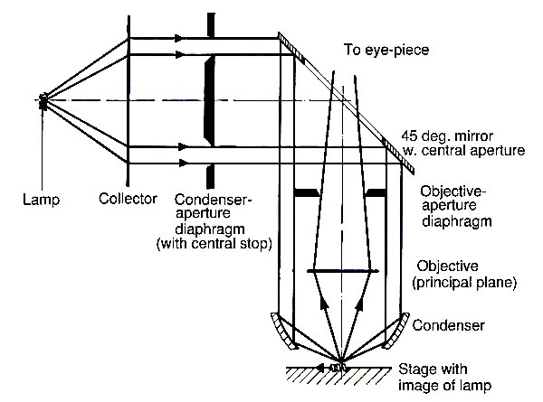
Dark Field (DF)
The light is emanating from the illumination lamp and collimated by the Collector. Most of the light is then blocked by the Condenser-aperture-diaphragm, which has a central stop that blocks the central rays of the beam. The light is then reflected by a 45° mirror with a circular hole in the center. In this way the light travels towards the sample on the outside of the actual objective lens. It is then focused on the sample by an inner mirror at the end of the objective housing. Light is deflected by any unevenness on the sample surface, and part of it is passed back via the objective lens to the eye of the operator. In this way, no directly (specularly) reflected light is seen by the operator (i.e. a perfectly clean mirror surface appears black). Only height differences, given by the dust and edges, on the surface are seen in the image.
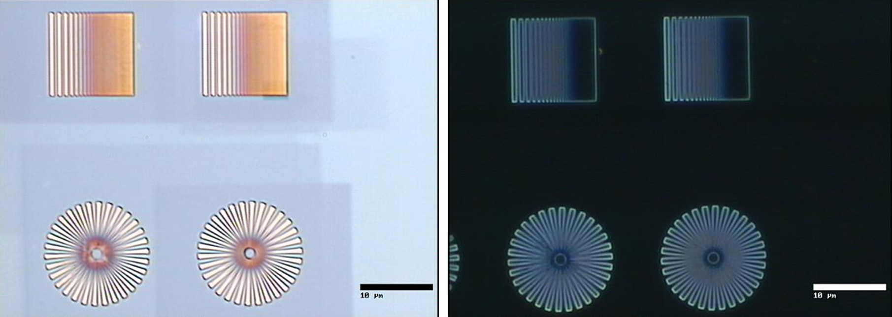
Bright field (left) and dark field (right) images of a test sample. In DF only the edges are visible.